![]() Figure 2 of
Nakamura, Mol Vis 2007;
13:1451-1457.
Figure 2 of
Nakamura, Mol Vis 2007;
13:1451-1457.
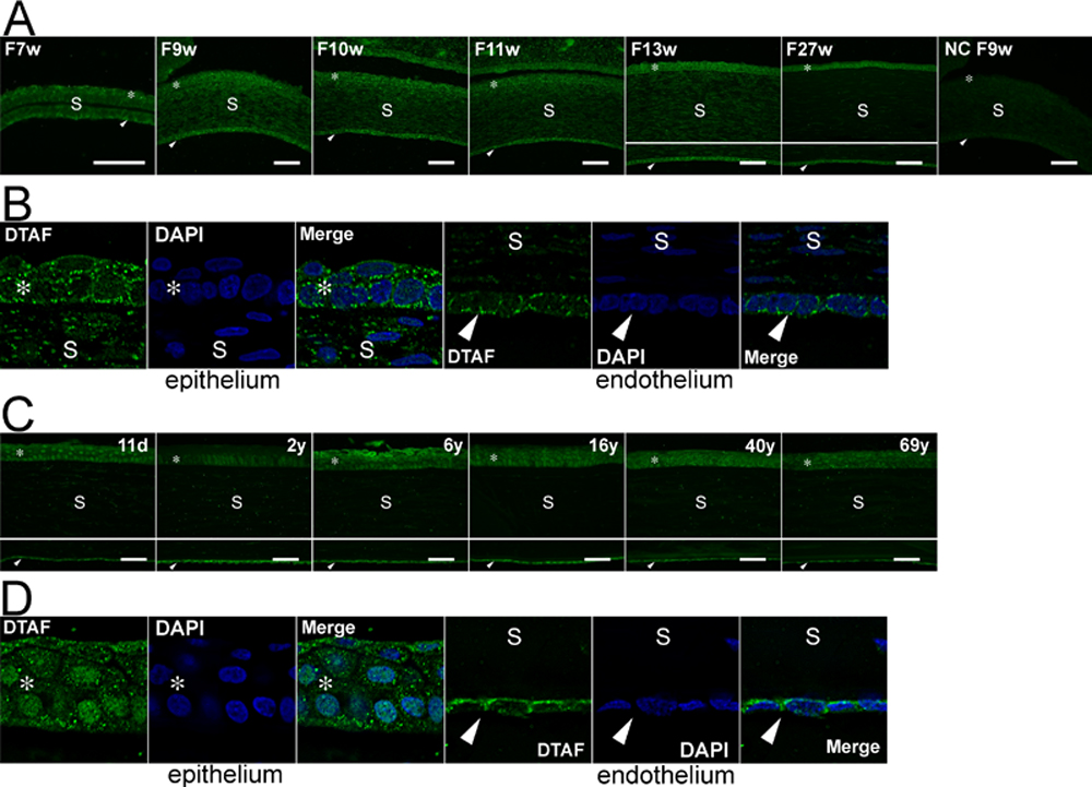
Figure 2. Immunofluorescence staining of KLF6 in the human cornea
The fluorescence staining for KLF6 and nucleus was visualized with DTAF (green) and DAPI (blue), respectively. A: Staining at different fetal stages. Positive KLF6 staining was noted in the specimen from a human fetus (F) of 7 weeks (w) in all corneal layers. In the epithelium and endothelium, the staining was mostly cytoplasmic at all the fetal stages studied. The intensity of KLF6 staining in the epithelium and endothelium remained relatively constant throughout fetal stages. The KLF6 staining in the stroma however was reduced at F27w. B: Deconvolution images at F13w. KLF6 immunostaining was obvious in the cytoplasm of epithelial and endothelial cells. C: Staining at new born to adult stages. After birth, the KLF6 staining appeared in the nuclei of corneal epithelial cells along with that in the cytoplasm. The intensity of KLF6 staining in the epithelium and endothelium remained relatively constant throughout these stages. The KLF6 staining in the stroma was invisible. D: Deconvolution images of specimen from an 11-day (d)-old child. Nuclear KLF6 immunostaining was clearly seen. Asterisks and arrowheads indicate the corneal epithelium and endothelium, respectively. S, stroma. The scale bar is equal to 50 μm. NC, negative control. The nuclear staining in the epithelium at F11w and 11d was scored as ± and ++, respectively.