![]() Figure 2 of
Rezaie, Mol Vis 2007;
13:1446-1450.
Figure 2 of
Rezaie, Mol Vis 2007;
13:1446-1450.
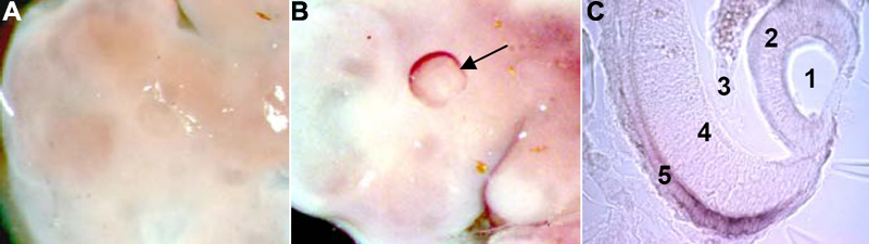
Figure 2. Higher resolution of Optn mRNA at age 10.5 dpc
A: Control and B: Optn probes. The arrow points to the prominent and specific expression of Optn in the developing eye. C: Frontal section of the developing eye at magnification 400X, cavity of the lens vesicle (1), lens vesicle (2), primary vitreous (3) neuroblastic retina (4), and outer layer of the optic cup (future pigment layer of the retina; 5).