![]() Figure 4 of
Shyong, Mol Vis 2007;
13:133-141.
Figure 4 of
Shyong, Mol Vis 2007;
13:133-141.
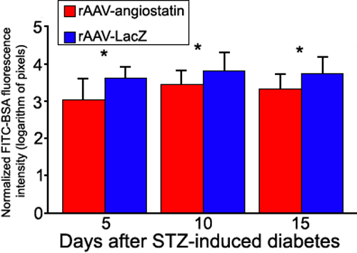
Figure 4. Quantification of vascular leakage in experimental diabetes
FITC-BSA fluorescence intensity was measured by image analysis in serial retinal sections. Rats each received an intravenous injection of FITC-BSA were sacrificed at 5, 10, and 15 days after induction of diabetes. The average retinal FITC-BSA fluorescence intensity was calculated and normalized to plasma fluorescence intensity. The retinal FITC-BSA fluorescence intensity in eyes receiving rAAV-angiostatin injection was 2.99±0.62 pixels at 5 days, 3.42±0.38 pixels at 10 days and 3.30±0.40 pixels at 15 days after induction of diabetes. The retinal FITC-BSA fluorescence intensity in eyes receiving rAAV-lacZ injection was 3.59±0.31 pixels at 5 days, 3.77±0.51 pixels at 10 days and 3.71±0.47 pixels at 15 days after induction of diabetes. The normalized FITC-BSA fluorescence intensity in eyes receiving rAAV-angiostatin was decreased as compared to eyes receiving rAAV-lacZ at 5 days (t=3.67, n=49, p=0.001), 10 days (t=3.94, n=51, p<0.001), and 15 days (t=3.52, n=56, p=0.001) after STZ-induction of diabetes. The asterisk indicates a p less than or equal to 0.001. Four SD rats were represented by the number of sections (n) to be examined.