![]() Figure 1 of
Shyong, Mol Vis 2007;
13:133-141.
Figure 1 of
Shyong, Mol Vis 2007;
13:133-141.
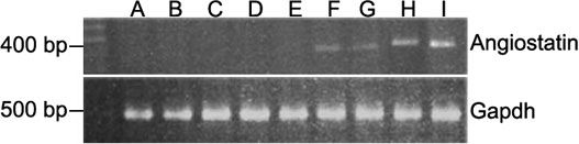
Figure 1. RT-PCR analysis of angiostatin cDNA in chorioretinal tissue
The eyes previously injected with rAAV-lacZ (lanes B to E) and rAAV-angiostatin (lanes F to I) were enucleated and chorioretinal tissues were harvested for RT-PCR at 1, 5, 10, and 15 days after induction of experimental diabetes. Lane A is the control eye. Lanes B and F are 1 day after diabetes induction. Lanes C and G are 5 days after diabetes induction. Lanes D and H are 10 days after diabetes induction. Lanes E and I are 15 days after diabetes induction. There was no angiostatin gene expression in the control eye (lane A) and eyes injected with rAAV-lacZ (lanes B to E). In the eyes injected with rAAV-angiostatin, angiostatin gene expression was detected (lanes F to I). As an internal control, expression of GAPDH was detected in normal control eye and eyes receiving both rAAV-angiostatin and rAAV-lacZ injections (lanes A to I). "M" indicates molecular weight markers.