![]() Figure 1 of
Alavi, Mol Vis 2007;
13:1441-1445.
Figure 1 of
Alavi, Mol Vis 2007;
13:1441-1445.
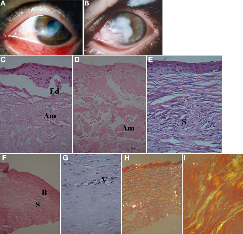
Figure 1. Slit lamp and histological appearance of the corneas
A,B: Slit lamp photographs of eye of, respectively, female and male GDLD patients. C-G: hematoxylin-eosin stained sections viewed under Tungsten filter; Ed: subepithelial edema, Am: staining suggestive of amyloid deposition, S: fibroblastic scars, B: disruption of Bowman's membrane, V: vascularization. H,I: congo red stained sections seen under polarized light; apple-green birefringence indicative of amyloid is present in section prepared after first (H) and second (I) surgical interventions. Histologic sections prepared from corneal tissue of male and female affected siblings were similar and the best are selected for presentation.