![]() Figure 5 of
Okada, Mol Vis 2007;
13:1428-1435.
Figure 5 of
Okada, Mol Vis 2007;
13:1428-1435.
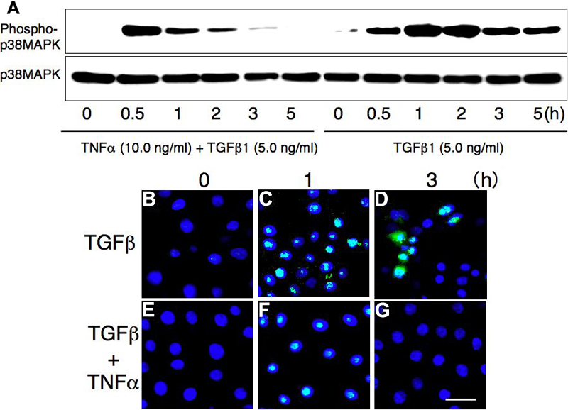
Figure 5. Effect of TNFα on TGFβ1-driven phosphorylated p38 mitogen-activated protein kinase (MAPK) in the Araki-Sasaki-corneal epithelial cell line
A: Bands for p38 (43 kDa) and phospho-p38 were observed in the cell lysate. TGFβ1 upregulates phosphorylation of p38MAPK with a peak at 1.0 h post-addition. Copresence of exogenous TNFα reduces the level of phosphorylation of p38MAPK at and after 1.0 h. At 5 h post-ligand(s) addition, phosph-p38MAPK is readily observed in the TGFβ1 culture, while not in the TGFβ1 plus TNFα culture. B-G: Immunocytochemistry shows that nuclear accumulation of phospho-p38MAPK is observed in both TGFβ1 or TGFβ1 plus TNFα cultures at 1 h, while it is seen in the TGFβ1 culture, but not in the TGFβ1 plus TNFα culture at 3 h (D versus G). The scale bar is equal to 30 μm.