![]() Figure 3 of
Okada, Mol Vis 2007;
13:1428-1435.
Figure 3 of
Okada, Mol Vis 2007;
13:1428-1435.
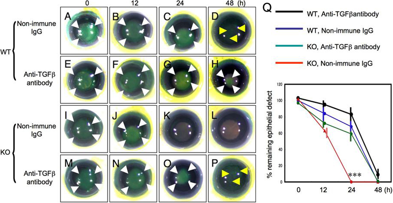
Figure 3. Reepithelialization of a circular epithelial debridement in an organ-cultured cornea from TNFα-knockout mice
A wild-type (WT, A-H) or TNFα-null (KO, I-P) mouse cornea with a circular epithelial defect was allowed to heal in culture medium supplemented with non-immune IgG (WT; A-D, KO; I-L) or anti-TGFβ neutralizing antibody (WT; E-H, KO; M-P). In WT cornea cultures, adding anti-TGFβ antibody retards reepothelialization. At 48 h a WT cornea exhibits a small punctate fluorescein staining without a definitive epithelial defect with non-immune IgG, while a circular defect remains with anti-TGF beta neutralizing antibody. The defect in a KO cornea reepithelialized in 24 h in a culture with non-immune IgG, indicating that loss of TNFα promotes reepithelialization in organ culture. Such promotion of epithelial healing in a KO cornea is reversed by adding anti-TGFβ neutralizing antibody to the medium. White arrowheads indicate a definitive epithelial defect and yellow arrowheads show punctate fluorescein staining. Q shows the percentage of remaining defect in each cornea. A cornea with punctate epithelial staining with fluorescein dye was grouped as a cornea without an epithelial defect. The bar in the graph (Q) is standard deviation.