![]() Figure 2 of
Okada, Mol Vis 2007;
13:1428-1435.
Figure 2 of
Okada, Mol Vis 2007;
13:1428-1435.
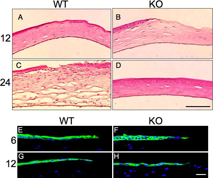
Figure 2. Histology and phospho-p38 mitogen-activated protein kinase (p38MAPK) expression
A-D: Histology of epithelium-debrided corneas stained with hematoxylin and eosin. No histological differences were seen in the healing corneas of the wild-type (WT; A) and TNFα-null (KO; B) mice at 12 h post-injury. However, at 24 h inflammation and stromal edema are observed in a WT cornea (C), while a KO cornea is reepithelialized with minimal inflammation in the stroma (D). E-H: Expression of phospho-p38MAPK is similar in migrating corneal epithelium between a WT (E,G) and KO (F,H) corneas at 6 and 22 h. The scale bar in D is equal to 50 μm and in H is equal to 10 μm.