![]() Figure 1 of
Okada, Mol Vis 2007;
13:1428-1435.
Figure 1 of
Okada, Mol Vis 2007;
13:1428-1435.
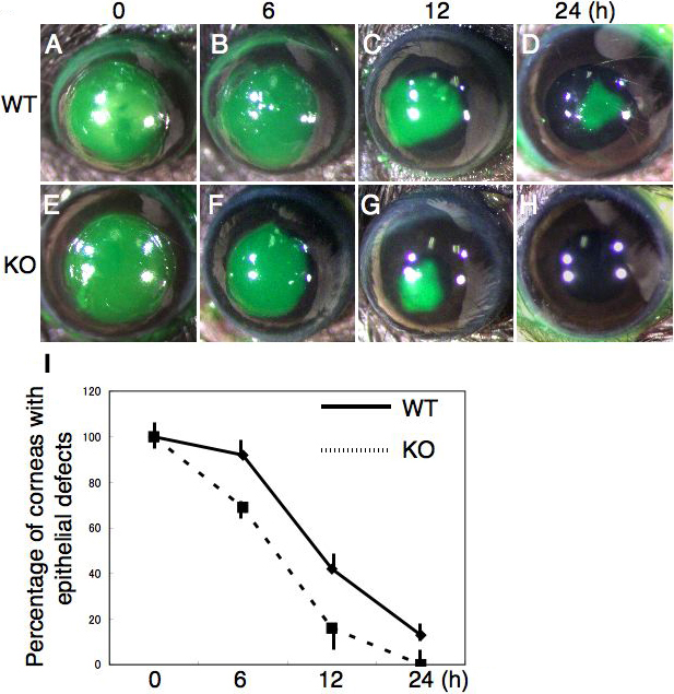
Figure 1. Reepithelialization of a circular epithelial debridement in a cornea from TNFα-knockout mice
This figure shows corneas of wild-type (WT; A-D) and TNFα-null (KO; E-H) mice at 0, 6, 12, and 24 h post-debridement, respectively. Remaining epithelial defects are stained with green fluorescein. At 6 and 12 h, defects are smaller in KO mice (F,G) as compared with WT corneas (B,C). At 24 h, a KO cornea is reepithelialized (H), while a WT cornea exhibits a remaining defect (D). I: Incidence (%) of the eyes with epithelial defects at each timepoint. Bar, standard deviation.