![]() Figure 9 of
Raji, Mol Vis 2007;
13:1412-1427.
Figure 9 of
Raji, Mol Vis 2007;
13:1412-1427.
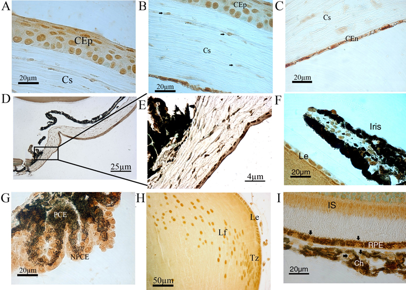
Figure 9. Msi-1 protein production in non-retinal tissues
High magnification of the cornea shows intense immunolabeling of the corneal epithelium (CEp; A), the corneal keratinocytes (A and B), and the corneal endothelial cells (CEn; C). D and E: Msi1 was observed in the limbal cells including the stem and progenitor cells. F: Msi1 immunoreactivity was also detected in the two pigmented layers of the iris and in iris stromal melanocytes and iris endothelial cells. G: Msi1 immunoreactivity was also detected in the pigmented ciliary epithelium (PCE) and nonpigmented ciliary epithelium (NPCE) of the ciliary body. H: Msi1 was observed in the lens epithelium (Le), transitional zone (Tz), and lens fiber (Lf). I: Significant immunoreactivity was observed in the retinal pigment epithelium (RPE) and the inner segment (IS).