![]() Figure 7 of
Raji, Mol Vis 2007;
13:1412-1427.
Figure 7 of
Raji, Mol Vis 2007;
13:1412-1427.
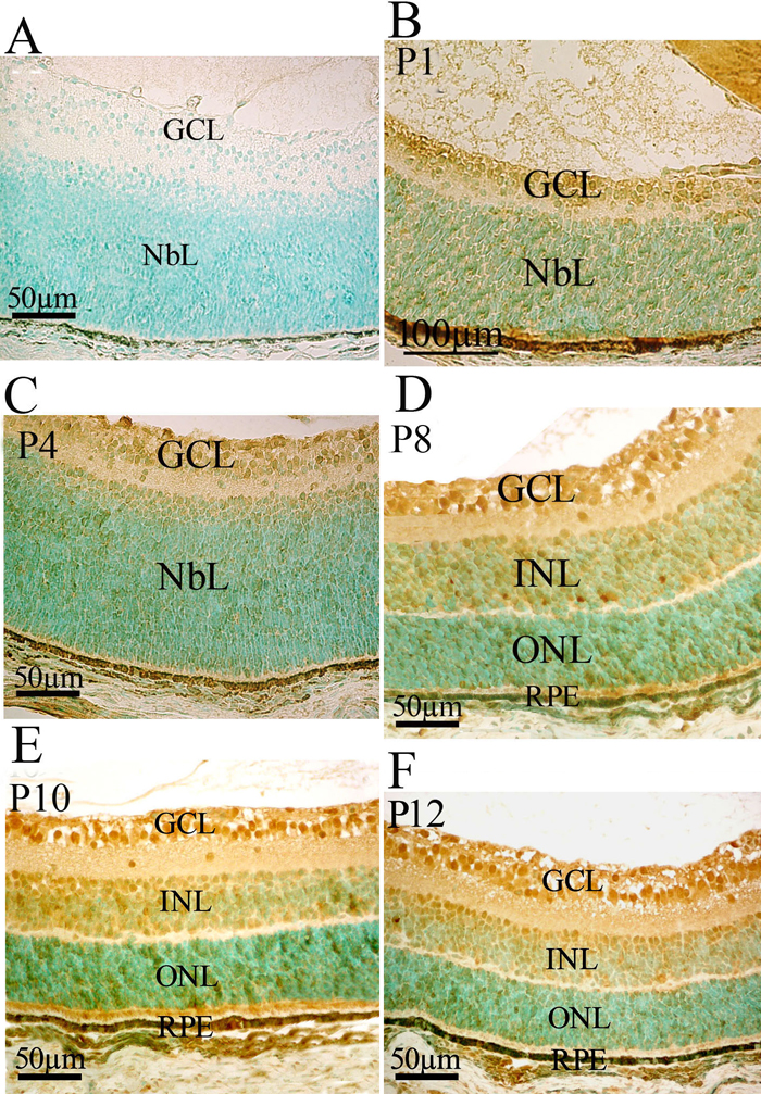
Figure 7. Msi1 protein production in the developing mouse retina
Protein was detected by immunochemistry, using a rabbit anti-mouse Msi1 antibody with 3% methyl green counterstaining. Msi1 is present principally in the ganglion cell layer (GCL) and in the outer region of the neuroblastic layer (NbL) of the developing retina (B and C). This pattern persists throughout postnatal stages of development with the intensity of Msi1 immunolabeling in the GCL increasing in later stages of development (D-F). Msi1 is dispersed in the INL and ONL late in retinal development (D-F). A: Immunonegative control. NbL, neuroblastic layer; GCL, ganglion cell layer; INL, inner nuclear layer; ONL, outer nuclear layer; RPE, retinal pigment epithelium.