![]() Figure 5 of
Raji, Mol Vis 2007;
13:1412-1427.
Figure 5 of
Raji, Mol Vis 2007;
13:1412-1427.
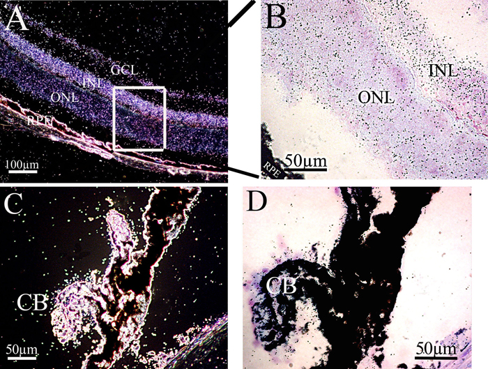
Figure 5. Msi1 mRNA production in the adult eye
A: In adults, Msi1 mRNA was still detected in the neuroretina with a strong signal observed in the ganglion cell layer, inner nuclear layer, and outer nuclear layer. B: Histological appearance of the same retina observed in bright field at high magnification, making it possible to distinguish the black grains corresponding to labeling. RPE, retinal pigmented epithelium; GCL, ganglion cell layer; INL, inner nuclear layer; ONL, outer nuclear layer; C-D: Msi1 mRNA was also detected in the ciliary body (CB) at the adult stage. The image in D corresponds to that in C in bright field at high magnification, making it possible to distinguish the black grains corresponding to labeling.