![]() Figure 1 of
Raji, Mol Vis 2007;
13:1412-1427.
Figure 1 of
Raji, Mol Vis 2007;
13:1412-1427.
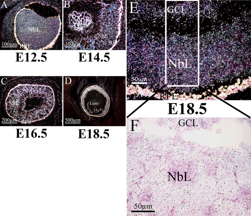
Figure 1. Msi1 mRNA production in embryonic mouse eye
A-D: In the retinal labeling observed in a dark field, Msi1 transcripts were detected at high levels in the neuroblastic layers of the retina from E12.5 to E18.5. E-F: A high magnification confirms the presence of Msi1 labeling in the retina in (E) the dark-field image (white grains) and in (F) the bright-field image (black grains). NbL, neuroblastic layer; GCL, ganglion cell layer; RPE, retinal pigment epithelium; Le, lens epithelium cells; Lf, lens fiber, Tz, transitional zone.