![]() Figure 3 of
Mulhern, Mol Vis 2007;
13:1397-1405.
Figure 3 of
Mulhern, Mol Vis 2007;
13:1397-1405.
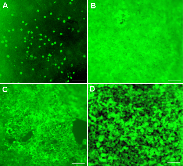
Figure 3. Lens epithelial cells from galactose-fed rat lenses stained with a calcein viability dye
Lenses from rats treated with PBS control (A) or the cellular osmolytes (PBA, B; TMAO, C; or TUDCA, D) were obtained after 15 days of galactose feeding. Lenses were isolated from the eyes, then whole lenses were stained with 0.5 ml of calcein for 40 min according to the manufacturer's protocol. The lenses were washed three times with PBS and photographed by fluorescent microscopy. All pictures were taken from the central region of the LECs where we can focus well. The scale bar indicates 100 μm.