![]() Figure 2 of
Blanco-Marchite, Mol Vis 2007;
13:1390-1396.
Figure 2 of
Blanco-Marchite, Mol Vis 2007;
13:1390-1396.
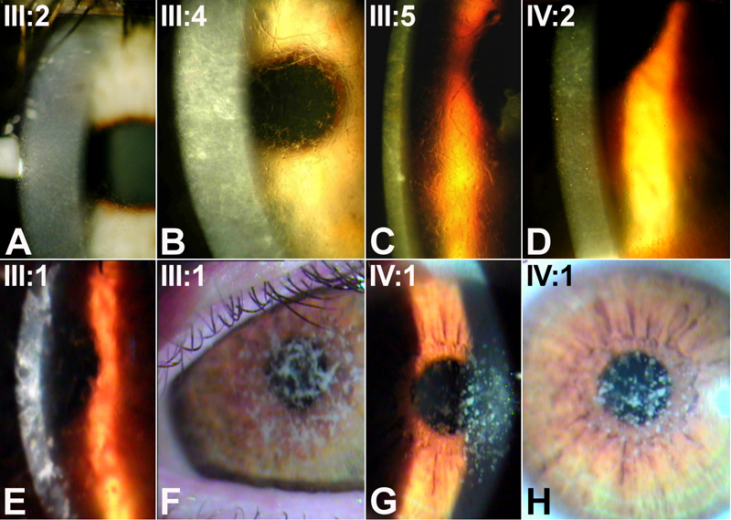
Figure 2. Corneal phenotypes as shown by slit lamp examination
Shown are photographs of lattice type I corneal dystrophy in family CD1 (A-D) and granular type I corneal dystrophy in family CD2 (E-H). The image in A corresponds to the transplanted cornea. Photographs A-D, F, and H were taken using retroillumination, while photographs E and G were obtained using direct illumination. The numbers correspond to individuals from pedigrees in Figure 1.