![]() Figure 8 of
Hu, Mol Vis 2007;
13:1379-1389.
Figure 8 of
Hu, Mol Vis 2007;
13:1379-1389.
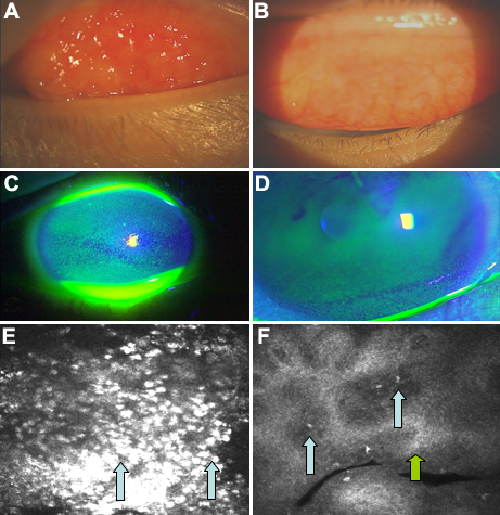
Figure 8. Pre- and post-treatment anterior segment photographs and confocal scan images from a patient with atopic keratoconjunctivitis using topical cyclosporine
A and B show the improvements in the injection and edema of the tarsal conjunctiva that is the regression of papillary formations in a patient with AKC (refractory to steroid and anti-allergic eye drop for eight weeks) who received eight weeks of topical cyclosporine drops in addition to the anti-allergic and steroid eye drops. C and D show the improvements in the corneal epithelial damage with cyclosporine treatment. E and F show the dramatic decrease in the conjunctival inflammatory cell infiltrates on the surface of the papillary formations (blue arrows) and the fibrotic response within the papillary formations (green arrow).
