![]() Figure 7 of
Hu, Mol Vis 2007;
13:1379-1389.
Figure 7 of
Hu, Mol Vis 2007;
13:1379-1389.
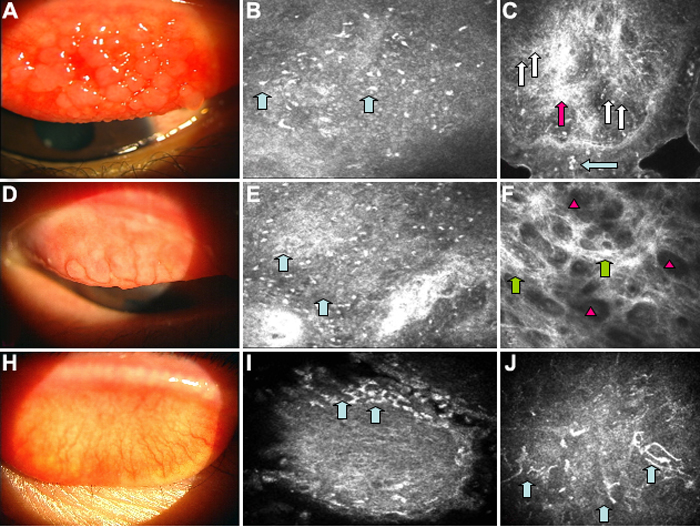
Figure 7. Conjunctival slit lamp photograph and confocal scan images from patients with atopic keratoconjunctivitis on topical cyclosporine
Upper row: A: The slit lamp photograph reveals injection of the tarsal conjunctiva and papillary hypertrophy. B: Confocal images demonstrated less inflammation on the surface of the papillary formations (blue arrows). C: Deeper images in the papillary formations showed fibrosis (pink arrow) and vascular neoformations (white arrows) with few inflammatory infiltrates (blue arrow). Middle row: This row shows the conjunctival slit lamp photograph (D) and confocal scan images from another patient with AKC on topical cyclosporine. E: Note the papillary hypertrophy and conjunctival hyperemia in the slit lamp photograph. Confocal images demonstrated less inflammation on the surface of the papillary formations (blue arrows). F: Deeper sections revealed extensive fibrosis(green arrows) with lacunar spaces displaying no inflammatory infiltrates (pink triangles). Lower row: Conjunctival slit lamp photograph and confocal scan images is shown from another patient with AKC. H: Note the conjunctival hyperemia in the slit lamp photograph. Confocal scans demonstrated lesser inflammation on the edges of the surface of the papillary formations (blue arrows; I) and dendritic cells (blue arrows; J).