![]() Figure 6 of
Hu, Mol Vis 2007;
13:1379-1389.
Figure 6 of
Hu, Mol Vis 2007;
13:1379-1389.
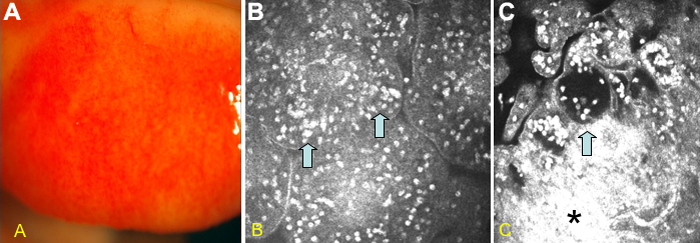
Figure 6. Conjunctival slit lamp photograph and confocal scan images from a patient with atopic keratoconjunctivitis on topical steroid and topical anti-allergic only
The slit lamp photograph reveals cherry red injection of the tarsal conjunctiva (A). Confocal images demonstrated extensive inflammation on the surface of the papillary formations (blue arrows; B). Deeper images in the papillary formations showed hyperreflective edematous areas (black asterisks) and lacunae with inflammatory infiltrates (blue arrow; C).