![]() Figure 5 of
Hu, Mol Vis 2007;
13:1379-1389.
Figure 5 of
Hu, Mol Vis 2007;
13:1379-1389.
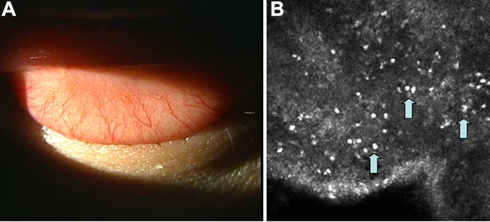
Figure 5. Conjuncival slit lamp photograph and confocal scan images from a normal control subject
Note the absence of papillary formations (A) and the few inflammatory cells dispersed in the conjunctical epithelium in the control subject (B). Note the absence of highly reflective areas suggesting fibrosis in the normal control eyes.