![]() Figure 1 of
Comes, Mol Vis 2007;
13:1363-1374.
Figure 1 of
Comes, Mol Vis 2007;
13:1363-1374.
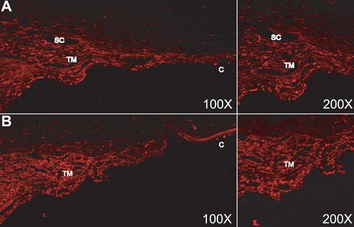
Figure 1. Fluorescence localization of labeled short-interfering RNA delivered to the intact human trabecular meshwork by intracameral perfusion
Post-mortem human anterior segments were perfused with Cy3 labeled siRNA for two days followed by perfusion with fresh DMEM for 24 h. After perfusion, tissues were fixed for one h in 4% paraformaldehyde, embedded in OCT and processed for histological evaluation. Confocal analysis of 10 μm sections from individual 1 (top) and individual 2 (bottom) showed high levels of fluorescence in the trabecular meshwork region. TM: trabecular meshwork, SC: Schlemm's Canal, C: cornea. Original magnification: 100X (left) and 200X (right).