![]() Figure 3 of
He, Mol Vis 2007;
13:1348-1356.
Figure 3 of
He, Mol Vis 2007;
13:1348-1356.
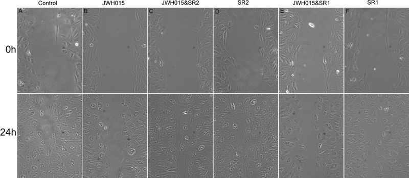
Figure 3. The effects of JWH015 on trabecular meshwork cell migration (wound healing)
TM cells grown to confluence were starved for 48 h and scratched with a yellow pipette tip to create a cell-free line. The medium was replaced with fresh medium containing vehicle (A), 100 nM JWH015 (B), 100 nM JWH015 plus 1 μM SR144528 (SR2; C), 1 μM of SR144528 (SR2; D), 100 nM JWH015 plus 1 μM SR141716A (SR1; E), or 1 μM SR141716A (SR1; F). The phase-contrast images (10X field) of the wound healing process were photographed digitally with an inverted microscope at 0 h (right after the scratching wound) and after incubation of cells at 37 °C for 24 h. The distances of the wound areas were measured on the images, set at 100% for 0 h, and the mean percentages of the total distances of the wound areas were calculated. The data are presented as mean ± S.E.M. Asterisks denote that data is significantly different from the vehicle control (p<0.05; one-way ANOVA with Neuman-Keuls post test). JWH015 caused a significant decrease in migration of TM cells to the wound area, compared to untreated control. This inhibitory effect of JWH015 was antagonized by SR144528 but not SR141716A.