![]() Figure 4 of
Matsumoto, Mol Vis 2007;
13:1319-1326.
Figure 4 of
Matsumoto, Mol Vis 2007;
13:1319-1326.
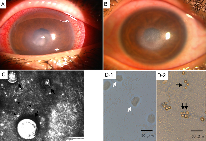
Figure 4. Clinical outcome, confocal scan and culture photographs of case 4
A: The pre-treatment slit-lamp photograph of the cornea revealed extensive subepithelial infiltration in the central cornea with conjunctival injection. B: The post-treatment slit-lamp photograph of the cornea revealed moderate remaining stromal opacity. C: The HRTII-RCM scan of the corneal lesion showed double-walled and round Acanthamoeba cysts (indicated by the black large arrows; upper left cyst size: 50 μm, upper right cyst: 40 μm, lower left cyst: 90 μm, depth: 62 μm) and small and round inflammatory cells (indicated by the black small arrow heads). D: Note that the presence of the Acanthamoeba trophozoites (indicated by the white arrows) was also confirmed by culture of corneal scrapings (D-1). Cysts (indicated by the black arrows) were also confirmed in the SCL storage solution (D-2).