![]() Figure 3 of
Matsumoto, Mol Vis 2007;
13:1319-1326.
Figure 3 of
Matsumoto, Mol Vis 2007;
13:1319-1326.
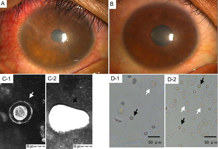
Figure 3. Clinical outcome, confocal scan and culture photographs of case 3
A: A slit-lamp photograph of the pre-treatment cornea revealed central subepithelial infiltration with radial keratoneuritis, corneal edema with conjunctival injection. B: A slit-lamp photograph of the post-treatment cornea revealed centrally clear cornea with mild nasal corneal opacity. C: The HRTII-RCM scan of the lesion showed double-walled and round Acanthamoeba cyst (C-1: indicated by the white arrow, size: 100 μm; depth: 2 μm) and highly reflective and irregular-shaped trophozoite (C-2: indicated by the black arrow, size: 175 μm; depth: 2 μm). D: Note that the Acanthamoeba cysts (indicated by the black arrow) and trophozoites (indicated by the white arrow) were reconfirmed by cultures of corneal scrapings (D-1). Cysts (indicated by the black arrows) and trophozoites (indicated by the white arrows) were also confirmed in the culture of the SCL storage solution (D-2).