![]() Figure 2 of
Matsumoto, Mol Vis 2007;
13:1319-1326.
Figure 2 of
Matsumoto, Mol Vis 2007;
13:1319-1326.
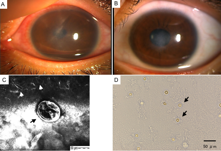
Figure 2. Clinical outcome, confocal scan and culture photographs of case 2
A: The pre-treatment slit-lamp photograph of cornea revealed subepithelial infiltration with radial keratoneuritis in the central cornea with conjunctival injection. B: The post-treatment slit-lamp photograph of the cornea revealed mild remaining corneal opacity. C: The HRTII-RCM showed a double-walled, round Acanthamoeba cyst (indicated by the black arrow; size: 100 μm; depth: 30 μm). Please note the Langerhans cells (indicated by the white arrows) in the close vicinity of the cyst. D: Note the Acanthamoeba cysts (indicated by the black arrows) confirmed by culture of soft contact lens (SCL) storage solution.