![]() Figure 1 of
Matsumoto, Mol Vis 2007;
13:1319-1326.
Figure 1 of
Matsumoto, Mol Vis 2007;
13:1319-1326.
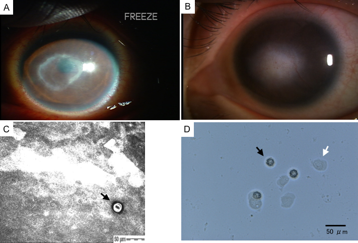
Figure 1. Clinical outcome, confocal scan, and culture photographs of case 1
A: The pre-treatment slit-lamp photograph of the cornea revealed conjunctival injection and advanced stage Acanthamoeba keratitis with ring infiltration in the central cornea. B: The post-treatment slit-lamp photograph of the cornea revealed round corneal opacity with deep new vessels in the central cornea. C: The HRTII-RCM showed a double-walled and round Acanthamoeba cyst (indicated by the black arrow; size: 25 μm; depth: 1 μm). D: Note the confirmation of the presence of the Acanthamoeba cysts (indicated by the black arrow) and trophozoites (indicated by the white arrow) in the culture of corneal scrapings.