![]() Figure 3 of
Agarwal, Mol Vis 2007;
13:1311-1318.
Figure 3 of
Agarwal, Mol Vis 2007;
13:1311-1318.
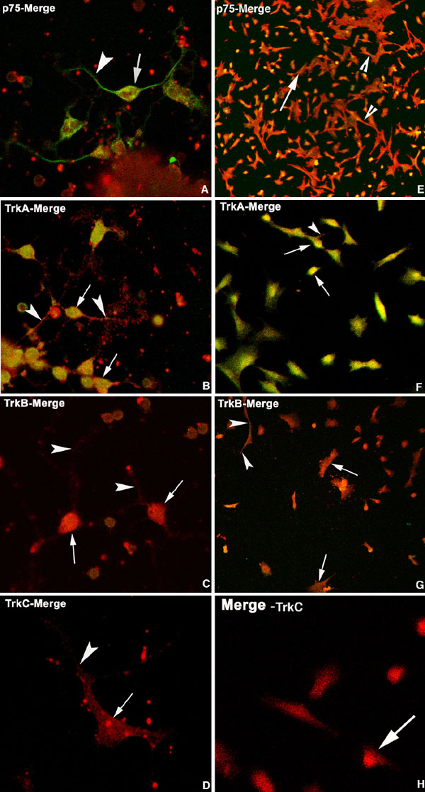
Figure 3. Confocal immunolocalization of neurotrophin receptors
Merged images of confocal immunocytochemical localization of p75 (green), TrkA (green), TrkB (green), and TrkC (green) and Thy-1 (red) in cultured adult RGCs (A-D, respectively) and retinal ganglion cell-5 (RGC-5) cells (E-H, respectively). Positive staining for p75 and TrkA with low levels of TrkB were observed in the soma (arrows) and neurites (arrowheads) of both cell types. Minimal if any, TrkC immunolabeling was detectable in either cell.