![]() Figure 1 of
Agarwal, Mol Vis 2007;
13:1311-1318.
Figure 1 of
Agarwal, Mol Vis 2007;
13:1311-1318.
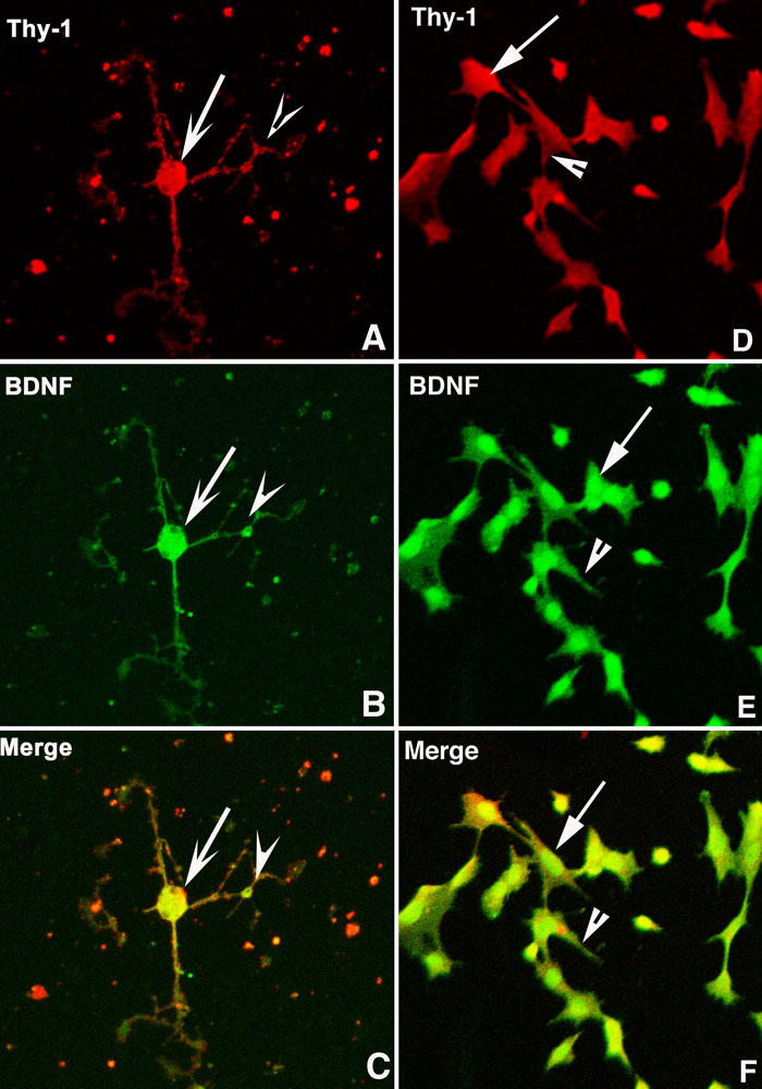
Figure 1. Confocal immunolocalization of brain-derived neurotrophic factor
Confocal immunocytochemical localization of Thy-1 (red) and brain-derived neurotrophic factor (BDNF; green) in primary culture of adult rat retinal ganglion cells (RGCs; A and B, respectively) and transformed RGC-5 cells (D and E, respectively). The merged images of these same cells are shown for primary (C) and transformed cells (F). BDNF and Thy-1 are co-expressed by both cells and are present in the soma (arrow) and the neurites (arrowhead).