![]() Figure 5 of
Zhou, Mol Vis 2007;
13:1298-1310.
Figure 5 of
Zhou, Mol Vis 2007;
13:1298-1310.
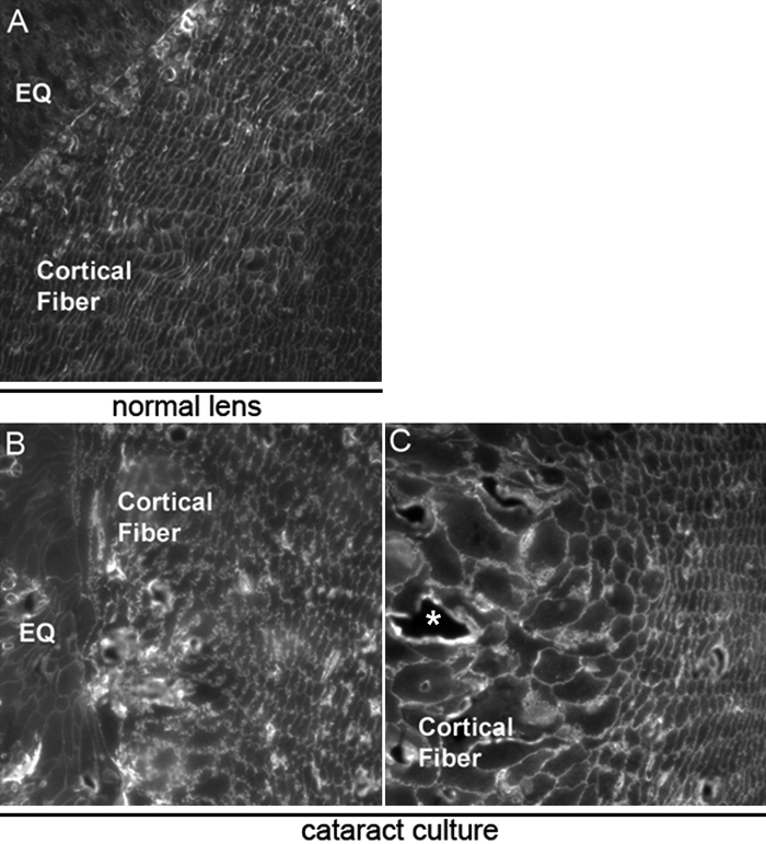
Figure 5. Dysmorphology of cortical fiber cells in cataract cultures
The highly organized structure and hexagonal packing of normal lens fiber cells in the cortical zone (A) is shown in a cross-section of the embryonic day 10 lens following immunostaining with an antibody to the major lens membrane intrinsic protein aquaporin-0 (MP28). The organization of cortical lens fiber cells and their membrane structure were also examined in cross-sections of lenses grown in cataract culture following immunostaining with antibody to aquaporin-0 (B,C). The highly organized hexagonal packing of the normal cortical fiber cells was disrupted in these cataractous lenses and individual cortical fiber cells were observed to have lost their linearity. Dysmorphology of the cortical fiber cells and separations between them (denoted by an asterisk) were common. EQ denotes equatorial zone.