![]() Figure 1 of
Zhou, Mol Vis 2007;
13:1298-1310.
Figure 1 of
Zhou, Mol Vis 2007;
13:1298-1310.
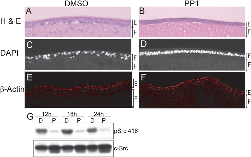
Figure 1. Disruption of the epithelium in cataract culture prevented by blocking activation of Src kinases
Lenses were grown in cataract culture conditions for ten days in the presence (B, D, F) and absence (A, C, E) of the Src kinase inhibitor PP1. Lens sections were observed by light microscopy following H&E staining (A, B) and by fluorescence microscopy following staining of nuclei with DAPI (C, D) and staining of actin filaments with fluorescent-conjugated phalloidin (E, F). Suppression of Src kinase activity by the PP1 inhibitor in the cataract cultures was confirmed by western blot analysis (G) at 12, 18, and 24 h in culture using the phosphoSrc418 (pSrc 418) antibody. Total Src expression was determined by western blot with Src antibody (c-Src). In the immunoblots, D (DMSO) denotes cataract culture and P the presence of the PP1 inhibitor. There was significant loss of integrity of the anterior epithelium in lenses grown in cataract culture that could be prevented by suppressing the activation of Src kinases. This included dysmorphology, multilayering, pycnotic and/or fragmented nuclei, and disorganization of the actin cytoskeleton. The epithelium in each photomicrograph is denoted by E and the fiber cell zone by F. Nuclei present in the anterior region of the fiber cell zone in the H&E stained section in A result from the aberrant organization of fiber cells that typically occurs in the cataract cultures (reported previously [20] and seen in the low power DAPI stained image in Figure 4B).