![]() Figure 1 of
Olofsson, Mol Vis 2007;
13:1285-1290.
Figure 1 of
Olofsson, Mol Vis 2007;
13:1285-1290.
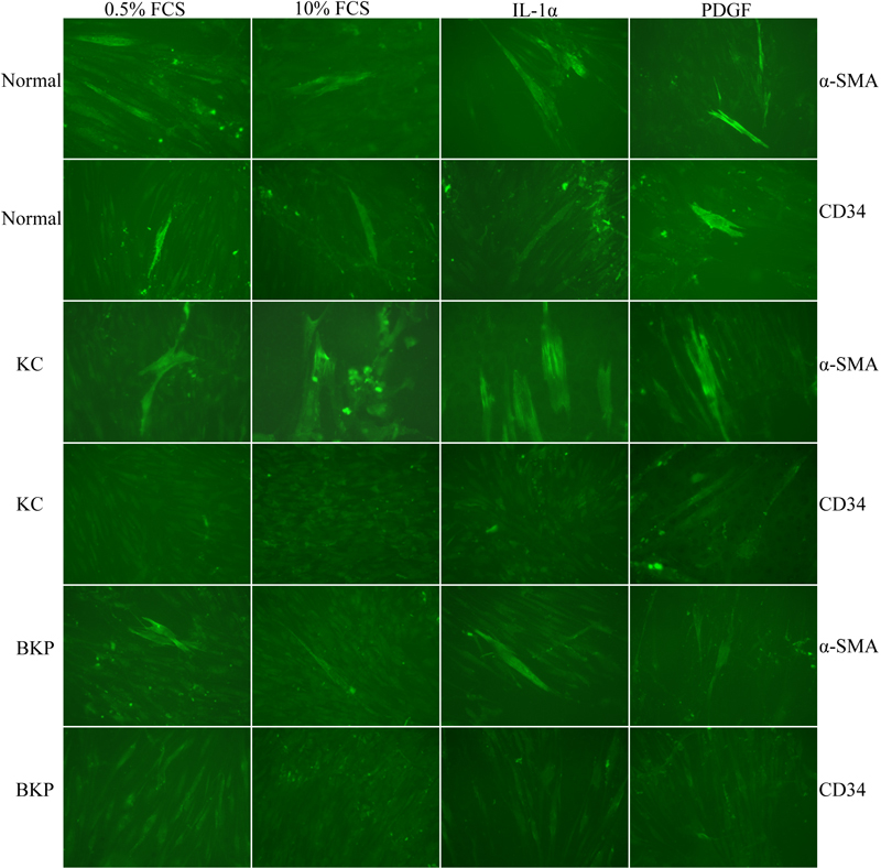
Figure 1. Cultured corneal stromal cells
Cultured stromal cells from a normal cornea, a cornea with keratoconus, and a cornea with bullous keratopathy were all stained with mouse monoclonal antibodies against α-SMA and CD34 after 96 h of culture in 0.5% or 10% FCS. The vast majority of the cells were unlabeled by the two antibodies. A few cells were labeled with the α-SMA antibody and even fewer with the CD34 antibody. No differences were seen between groups or treatments. The staining pattern suggests that the cells were of a fibroblast phenotype.