![]() Figure 6 of
Vasiliev, Mol Vis 2007;
13:114-124.
Figure 6 of
Vasiliev, Mol Vis 2007;
13:114-124.
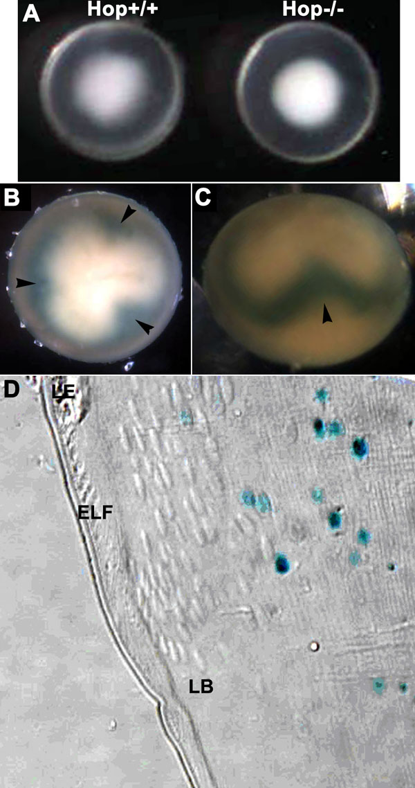
Figure 6. Appearance of Hop knockout lenses
Hop is expressed in maturing secondary fiber cells in the mouse lens. A: Hop wild type and null lenses from P3 mice. Both lenses have cold cataracts, as expected of lenses at this age. No consistent variations were detected in the size of the wild type and knockout lenses or in the extent of the cold cataracts. B: Polar view of a whole, Hop null lens stained for β-galactosidase activity. The superficial zone of the lens has no stained nuclei. The "trefoil" pattern of stained nuclei in the deeper fiber cells is due to the displacement of the nuclei in a more anterior or posterior direction as a result of differences in the extension of the fiber cells toward the anterior and posterior sutures [58]. C: The displacement of β-galactosidase-stained nuclei as viewed from the lens equator. D: A 1 μm plastic section of the equatorial region of a mouse lens in which both Hop alleles were disrupted by the insertion of the sequence encoding nuclear-targeted β-galactosidase [22]. The lens was stained for β-galactosidase activity, embedded in glycol methacrylate, and sectioned. The section was viewed using differential interference contrast optics to show the location of the nuclei of the fiber cells. Only the nuclei of the deeper fiber cells are stained blue, indicating that Hop expression is initiated late in fiber cell maturation. β-Galactosidase activity was still present in the fragments of nuclei remaining after organelle loss. The morphology of the cells of the Hop knockout lenses appears similar to wild type.