![]() Figure 5 of
Vasiliev, Mol Vis 2007;
13:114-124.
Figure 5 of
Vasiliev, Mol Vis 2007;
13:114-124.
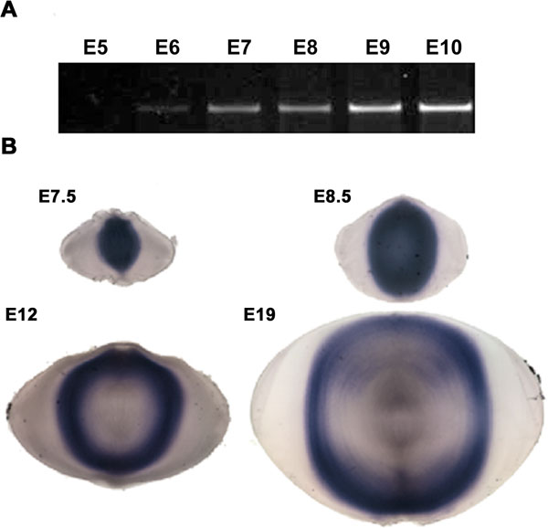
Figure 5. Hop expression during lens development
A: PCR amplification of Hop sequences in RNA extracted from chicken lenses from E5 through E10. Hop transcripts were first detectable at E6. Transcript levels increased at later stages. B: Hop is expressed soon after primary and secondary fiber cells detach from the capsule. In situ hybridization showing the distribution of Hop transcripts during lens development in chicken embryos. Sections are from lenses at E7.5, E8.5, E12, and E19. The decreased staining in the center of lenses at E12 and E19 probably reflects a decrease in probe penetration, not a decrease in Hop transcripts, because PCR analysis of microdissected lens cores from lenses at these stages revealed no obvious decrease in Hop sequences.