![]() Figure 2 of
Vasiliev, Mol Vis 2007;
13:114-124.
Figure 2 of
Vasiliev, Mol Vis 2007;
13:114-124.
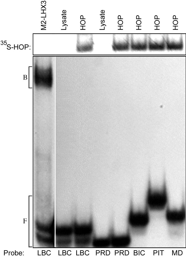
Figure 2. Hop does not bind to homeobox sequences
Electrophoretic mobility shift assay using radiolabeled oligonucleotide probes representing transcription factor binding sites. Probes were incubated with the indicated 35S-labeled in vitro translated proteins, and the bound complexes (B) were separated from the free probe (F) by electrophoresis. Unprogrammed lysate was used as a negative control (lysate). Bacterially expressed M2-LHX3 was used as a positive control [37]. Abbreviations: LBC=LHX3 LIM-class homeodomain site, PRD=paired-class homeodomain site, BIC=bicoid-class homeodomain site, PIT=Pit-1 POU-class homeodomain site, MD=MyoD basic helix-loop-helix site. The upper panel shows the input Hop protein (35S-labeled); the lower panel shows the migration of 32P-labeled DNA.