![]() Figure 3 of
DElia, Mol Vis 2007;
13:1245-1250.
Figure 3 of
DElia, Mol Vis 2007;
13:1245-1250.
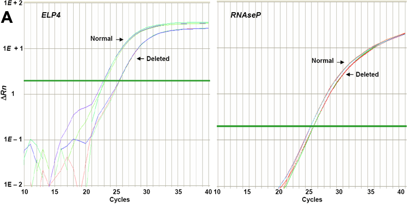
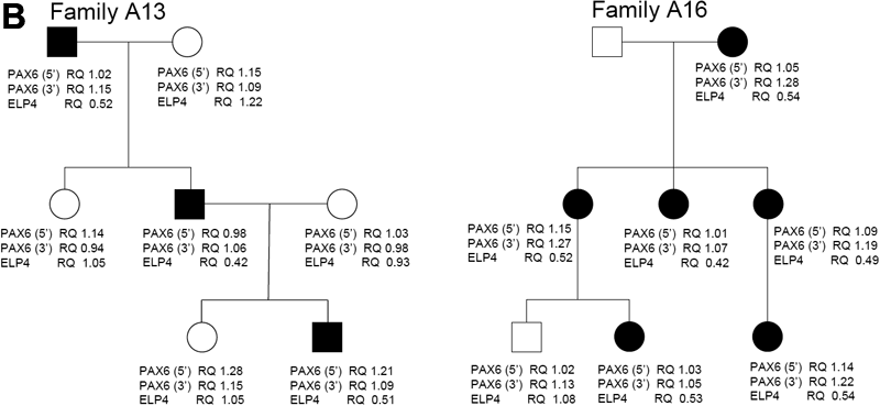
Figure 3. Analysis of deletions in the ELP4 gene region by real-time PCR
(A)This is an example of a fluorescence amplification plot of the quantitative real-time PCR for the ELP4 gene region. Arrows indicate triplicate signals obtained for a control subject (Normal) and an aniridia patient (Deleted). (B) Pedigrees of families with subjects having the ELP4 gene deletion are illustrated below. Below each subject, RQ values for the 5' and 3' PAX6 gene region as well as for the ELP4 gene region are indicated.