![]() Figure 8 of
Qiong, Mol Vis 2007;
13:1234-1244.
Figure 8 of
Qiong, Mol Vis 2007;
13:1234-1244.
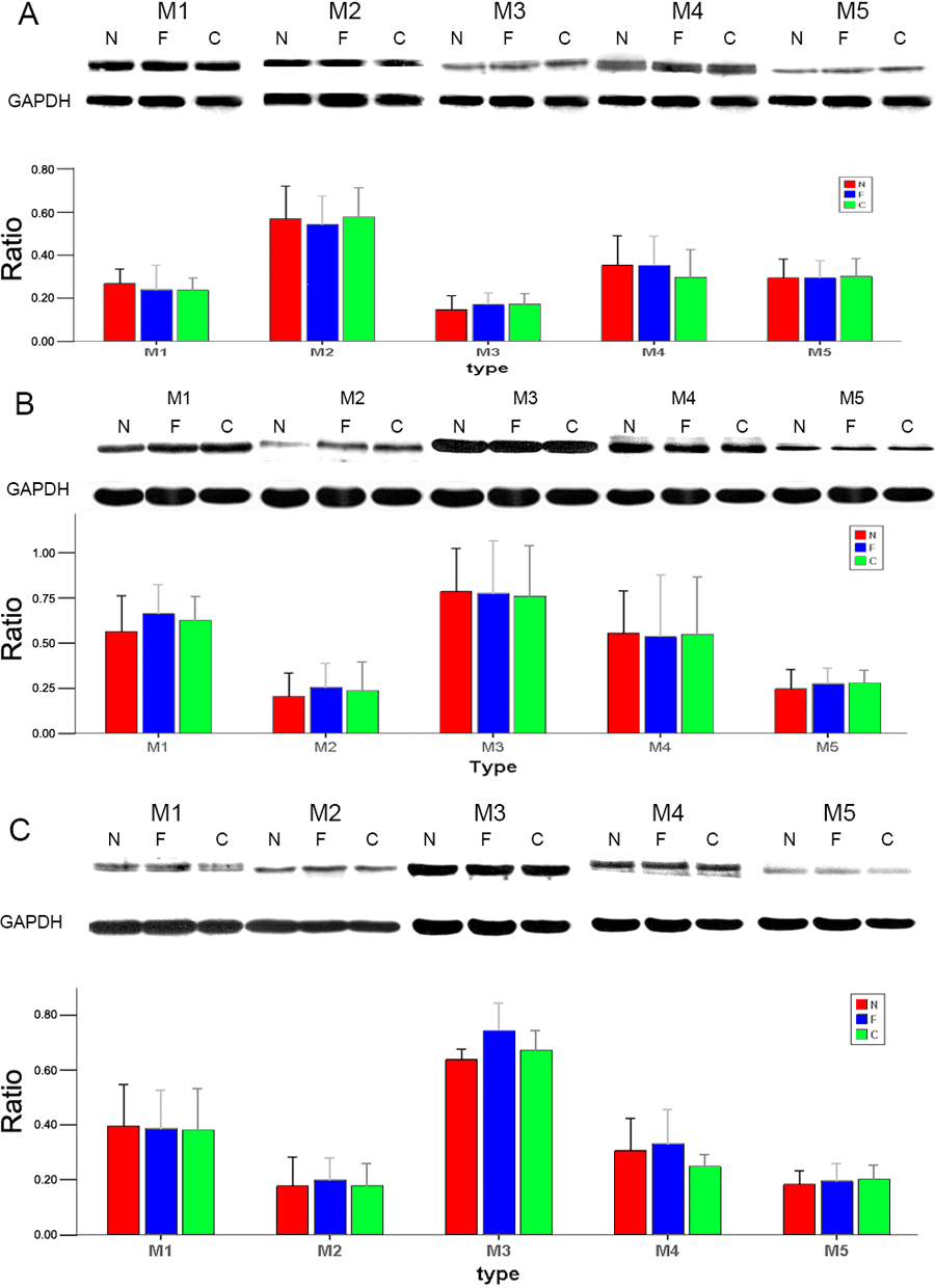
Figure 8. Changes in protein expression in the retina, choroid, and iris-ciliary body of guinea pigs
There were changes in protein expression in the retina (A), choroid (B), and iris-ciliary body (C) of guinea pigs. Typical gels indicate the level of GAPDH protein relative to those of receptor subtypes M1 to M5. Bar graph show changes in protein expression where values (means±standard error of the mean) were normalized for GAPDH and expressed as ratios of optical density. Semiquantitative western blotting showed no significant change for M1 to M5 subtype protein expression in form-deprived myopia (F) versus internal control (C) and normal (N) eyes.