![]() Figure 4 of
Qiong, Mol Vis 2007;
13:1234-1244.
Figure 4 of
Qiong, Mol Vis 2007;
13:1234-1244.
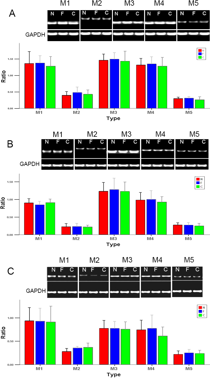
Figure 4. Changes in mRNA expression in the retina, choroid, and iris-ciliary body of guinea pigs after induction of form-deprived myopia
Changes in mRNA expression in the retina (A), choroid (B), and iris-ciliary body (C) of guinea pigs after induction of form-deprived myopia. Ethidium-bromide agarose gels indicate the level of glyceraldehyde-3-phosphate dehydrogenase (GAPDH) message relative to levels for receptor subtypes M1 to M5 from total RNA. Bar graphs show changes in mRNA expression (mean±standard error of the mean) where values were normalized to GAPDH and expressed as ratio of optical density. Semiquantitative reverse-transcription polymerase chain reaction showed no significant change for M1 to M5 in FDM (F) compared with the internal control (C) and normal (N) eyes.