![]() Figure 3 of
Qiong, Mol Vis 2007;
13:1234-1244.
Figure 3 of
Qiong, Mol Vis 2007;
13:1234-1244.
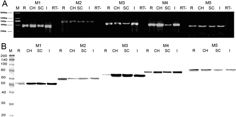
Figure 3. mRNA and protein expression for receptor subtypes M1 to M5 in the retina, choroid, sclera, and iris-ciliary body of normal guinea pigs
In A is gel electrophoresis analysis of polymerase chain reaction (PCR) products from the total RNA of the tissues. On RT-PCR, amplified products were about 437, 659, 506, 466, and 455 bp for the M1, M2, M3, M4, and M5 subtypes, respectively. The M1 to M5 subtypes were found in the retina, choroid, sclera, and iris-ciliary body. RT- represents the negative control, where PCR was run without an RNA template. B are the protein blots. Lanes were loaded with 40 μg of protein extracted from the various tissues. Proteins for the M1-M5 subtypes were present in normal ocular tissues. Molecular masses (left of each blot) indicate the positions of molecular-mass standards on the same gel. R: retina; CH: choroid; SC: sclera; I: iris-ciliary body.