![]() Figure 2 of
Wang, Mol Vis 2007;
13:1226-1233.
Figure 2 of
Wang, Mol Vis 2007;
13:1226-1233.
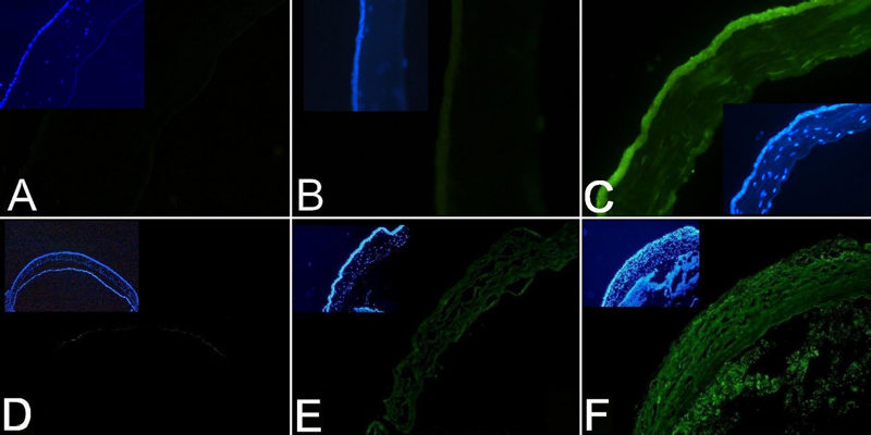
Figure 2. Immunohistochemistry detection of MBL-A in fungi-challenged murine corneas
Immunohistochemistry detection of MBL-A in in vitro-challenged corneas (A-C) and in corneas from in vivo experimental FK mice (D-F). For in vitro study, the corneas were either cultured with just the medium (A,B) or with heat-killed spores (C) for 24 h. FK was induced in Balb/c mice as described (F) and the corneas were removed three days later for IHC with sham-infected mice as the control (D,E). The specificity of staining was shown by the negative control (A,D, and data not shown) where sections from control corneas were handled in exactly the same way except that the primary antibody was omitted in the first staining step. Inlet in each panel shows DAPI staining of nuclei of the same section. Representative results from two (in vitro study) and three (in vivo study) experiments were shown. Capture modes used for pictures: DS-U1 CCD controlled with Nikon ACT-2U software (version 1.40.85) under fluorescence mode. Exposure control: Manual; Exposure time: 1/6 s.