![]() Figure 5 of
Makhani, Mol Vis 2007;
13:1215-1225.
Figure 5 of
Makhani, Mol Vis 2007;
13:1215-1225.
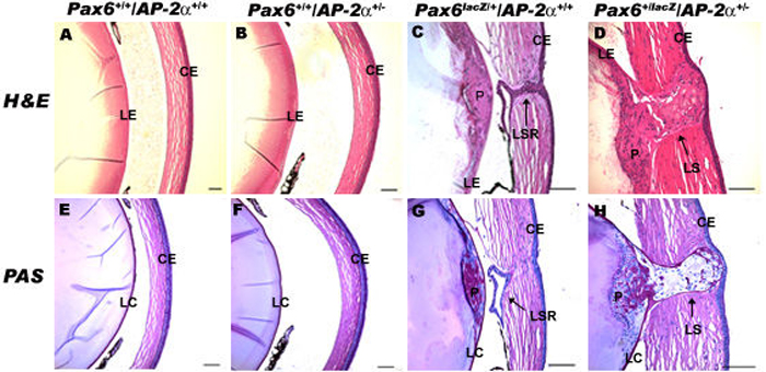
Figure 5. Pax6+/lacZ/AP-2α+/- adult mouse eyes
Histological examination of the adult eye sections were carried out using hematoxylin and eosin (H & E) staining as well as periodic acid schiff (PAS) staining. No defects were observed with H & E staining in the adult wild type eyes (A) and AP-2α+/- eyes (B). While both Pax6+/lacZ (C) and Pax6+/lacZ/AP-2α+/-(D) eyes demonstrated lens separation defects, only Pax6+/lacZ/AP-2α+/-eyes demonstrated a persistent lens stalk (D). Normal PAS staining was observed in adult wild type (E) and AP-2α+/- (F) eyes. PAS staining indicated matrix deposition within the plaques, the lens stalk remnants of Pax6+/lacZ (G), and the lens stalks of Pax6+/lacZ/AP-2α+/-(H) eyes. PAS staining also demonstrated continuity of the lens capsule around the stalk in the Pax6+/lacZ/AP-2α+/- eyes (H). LE, lens epithelium; CE, corneal epithelium; P, plaque; LSR, lens stalk remnant; LS, lens stalk; LC, lens capsule. All scale bars are 100 μm.