![]() Figure 3 of
Makhani, Mol Vis 2007;
13:1215-1225.
Figure 3 of
Makhani, Mol Vis 2007;
13:1215-1225.
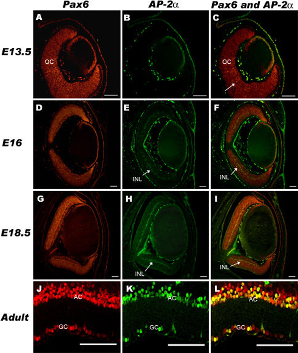
Figure 3. Colocalization of Pax6 and AP-2α in the developing and adult retina
Pax6 (red) and AP-2α (green) were colocalized (yellow) in a small subset of cells in the developing retina at E13.5 (A, B, C, the arrow points to a colocalized cell). Pax6 and AP-2α were colocalized in cells of the developing inner nuclear layer at E16 (D, E, F) and E18.5 (G, H, I). Pax6 and AP-2α were colocalized in amacrine cells of the inner nuclear layer and ganglion cell layer of the adult retina (J, K, L). Pax6 was detected with a rhodamine-conjugated secondary antibody and AP-2α was detected with a FITC-conjugated secondary antibody. OC, optic cup; INL, inner nuclear layer; AC, amacrine cells; GC, ganglion cells. All scale bars are 100 μm.