![]() Figure 1 of
Makhani, Mol Vis 2007;
13:1215-1225.
Figure 1 of
Makhani, Mol Vis 2007;
13:1215-1225.
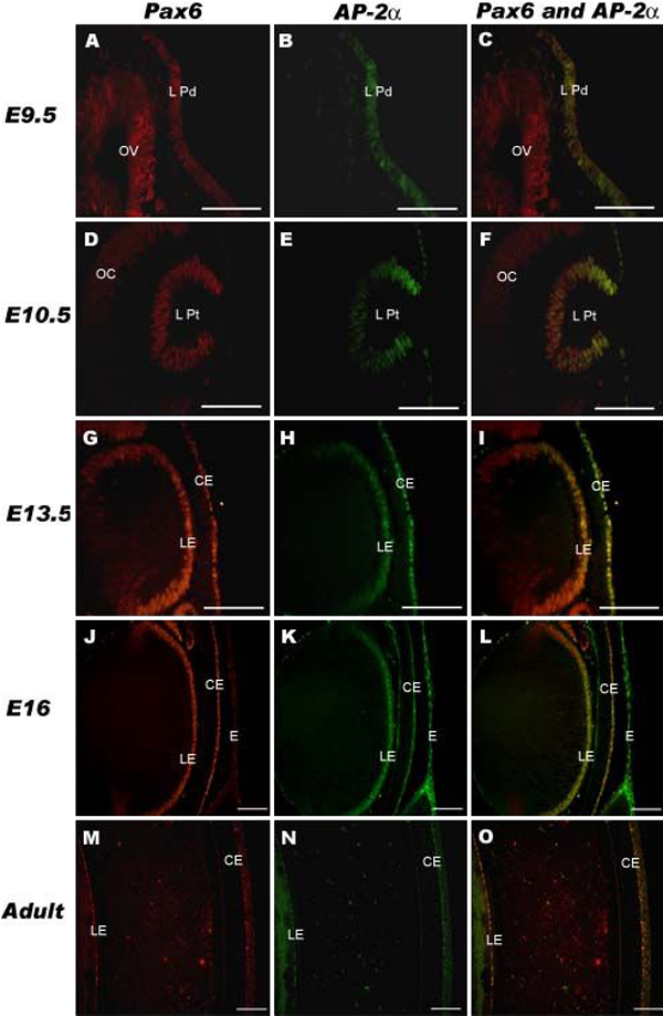
Figure 1. Colocalization of Pax6 and AP-2α in the developing lens epithelium
Pax6 (red) and AP-2α (green) expression was examined at E9.5, E10.5, E13.5, E16, and at the adult stage using immunofluorescence (C, F, I, L, O). Colocalization (yellow) was observed in the lens placode at E9.5 (C), in cells lining the lens pit at E10.5 (F), in the corneal and central lens epithelium at E13.5, E16, and post-natally (I, L, O), and in the epidermis at E16 (L). Pax6 was detected with a rhodamine-conjugated secondary antibody (A, D, G, J, M) and AP-2α was detected with a FITC-conjugated secondary antibody (B, E, H, K, N). OV, optic vesicle; L Pd, lens placode; OC, optic cup; L Pt, lens pit; LE, lens epithelium; CE, corneal epithelium; E, epidermis. All scale bars are 100 μm.