![]() Figure 9 of
Lin, Mol Vis 2007;
13:1203-1214.
Figure 9 of
Lin, Mol Vis 2007;
13:1203-1214.
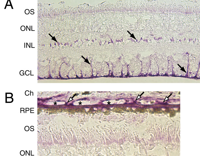
Figure 9. Immunohistochemical localization of the retinal G protein-coupled receptor splice isoform epitope in Müller cells
A: The retina section was prepared from the macular region of donor 960105, a 69-year-old male with macular degeneration, and incubated with the DE21 antibody. Positive staining (solid arrows) in the neural retina was evident in the somata and quite strongly, in the inner processes and endfeet of Müller cells. B: Higher magnification of positive staining of the intercapillary region (open arrows) and Bruch's membrane. Asterisks indicate lumen of the choriocapillaris. OS represents outer segments; ONL represents outer nuclear layer; INL represents inner nuclear layer; GCL represents ganglion cell layer; Ch represents choroid; RPE represents retina pigment epithelium.