![]() Figure 7 of
Lin, Mol Vis 2007;
13:1203-1214.
Figure 7 of
Lin, Mol Vis 2007;
13:1203-1214.
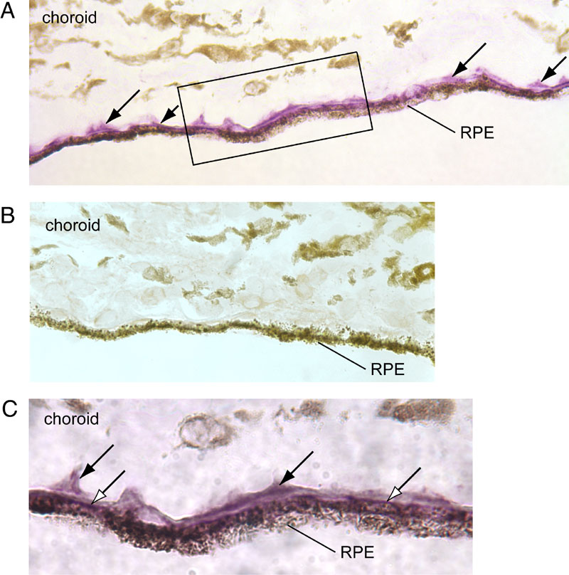
Figure 7. Immunohistochemical localization of the retinal G protein-coupled receptor splice isoform epitope in Bruch's membrane
A: The retina or retinal pigment epithelium (RPE)-choroid section was obtained from the macular region of donor Y-005 (OD) and incubated with the DE21 antibody. Arrows point to immunostaining with the VIP substrate in Bruch's membrane and intercapillary region of the choriocapillaris. B: Control RPE-choroid section incubated with peptide-blocked DE21 antibody. C: Higher magnification of boxed region in A, showing immunostaining of intercapillary regions (solid arrows) and basal boundary of RPE cells (open arrows). The neural retina was detached and absent from the section.