![]() Figure 3 of
Lin, Mol Vis 2007;
13:1203-1214.
Figure 3 of
Lin, Mol Vis 2007;
13:1203-1214.
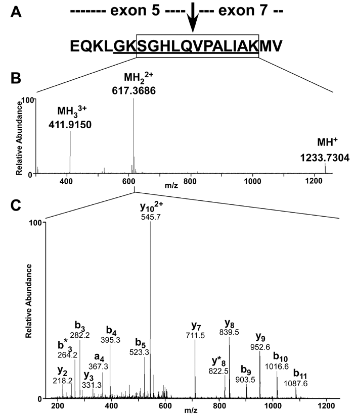
Figure 3. Mass spectrometric analysis of a unique retinal G protein-coupled receptor splice isoform splice-site peptide
A: Sequence of a test peptide that encompasses the splice junction site of exons 5 and 7 in retinal G protein-coupled receptor spilce isoform (RGR-d). The peptide contained a tryptic fragment (boxed sequence) and the RGR-d peptide epitope (underlined sequence) used to generate RGR-d specific antibodies. The test peptide was digested with trypsin and characterized by liquid chromatography-mass spectrometry (LC/MS/MS) analysis. B: High resolution mass spectrum for the RGR-d splice-site peptide, a tryptic fragment from the test peptide. Ions in the spectrum were annotated to show the charge state and the observed m/z values. C: fragment ion spectrum (MS/MS) spectrum for the RGR-d splice-site peptide. Prominent sequence-related ions in the spectrum are annotated to show the observed m/z value and fragment ion series assignment.