![]() Figure 2 of
Lin, Mol Vis 2007;
13:1203-1214.
Figure 2 of
Lin, Mol Vis 2007;
13:1203-1214.
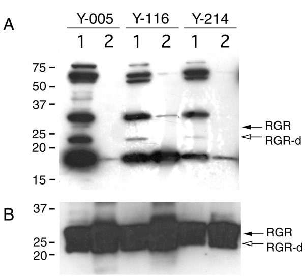
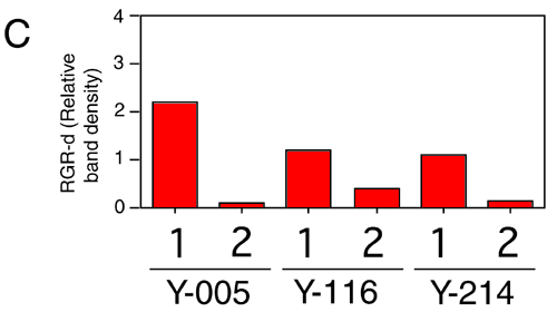
Figure 2. Western immunoblot assay of retinal G protein-coupled receptor splice isoform and retinal G protein-coupled receptor in the retina and retinal pigment epithelium of young donors
A single blot of the neural retina (lane 1) and retinal pigment epithelium (RPE; lane 2) membrane proteins from donors Y-005 (20/F), Y-116, (18/M), and Y-214 (21/M) was prepared so that retinal G protein-coupled receptor (RGR) would be about equal intensity in each lane. The blot was (A) incubated with the DE17 antibody, strip-washed, and then (B) incubated with the HRGR-DE7 antibody. C: The relative density of retinal G protein-coupled receptor splice isoform (RGR-d) bands was normalized to the band density of RGR. Monomer RGR-d (open arrows) and full-length RGR (solid arrows) were detected by chemiluminescence using (A) SuperSignal West Femto and (B) ECLTM substrate systems.