![]() Figure 7 of
Liang, Mol Vis 2007;
13:1169-1180.
Figure 7 of
Liang, Mol Vis 2007;
13:1169-1180.
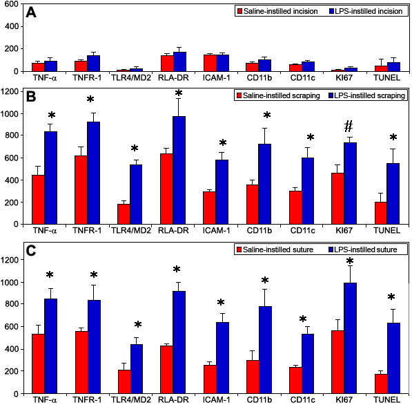
Figure 7. Illustrations of positive cell counts in the saline- or LPS-instilled corneal injury models
These are illustrations of positive cell counts for inflammatory, proliferative, and apoptotic markers in the saline- or LPS-instilled incision (A), scraping (B), and suture (C) models at D9 using anti-TNF-α, TNFR1, TLR4/MD2, RLA-DR, ICAM-1, CD11b, CD11c, Ki67 antibodies and TUNEL assay. The asterisk indicates that p<0.0001 and the sharp (hash mark) indicates that p<0.01 compared to the corresponding saline-instilled models.