![]() Figure 4 of
Liang, Mol Vis 2007;
13:1169-1180.
Figure 4 of
Liang, Mol Vis 2007;
13:1169-1180.
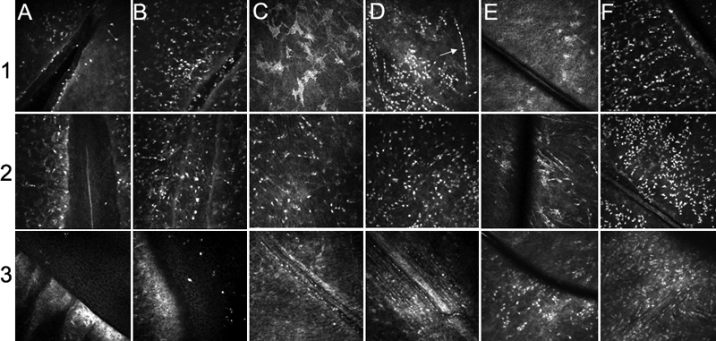
Figure 4. Heidelberg Retina tomography II in vivo confocal microscopy images in the saline- or LPS-instilled corneal injury models
Heidelberg Retina Tomograph II in vivo confocal microscopy images illustrates the results of the saline- or LPS-instilled incision models (A,B: 80-120 μm from the most superficial epithelial layer), of scraping models (C,D: 50-70 μm from the most superficial epithelial layer), and of suture models (E,F: 95-135 μm from the superficial epithelium layer) at H4 (line 1), D1 (line 2), and D9 (line 3). All LPS-receiving injury models (B,D,F) showed more inflammatory infiltration than saline-receiving models (A,C,E). In all LPS-receiving models (B,D,F), the inflammatory infiltrating cells were more abundant than in all saline-instilled models (A,C,E). Weakly reflective star-like cells in H4 (C) and H4 (E) are keratocytes whereas inflammatory elements, whatever their shape, appear as highly reflective cells. Corneal new blood vessels are clearly visible in scraping models (D9; C and D) and in suture models (D9; E andF). The black line in H4 E to D9 E and H4 F to D9 F correspond to nylon sutures.