![]() Figure 1 of
Liang, Mol Vis 2007;
13:1169-1180.
Figure 1 of
Liang, Mol Vis 2007;
13:1169-1180.
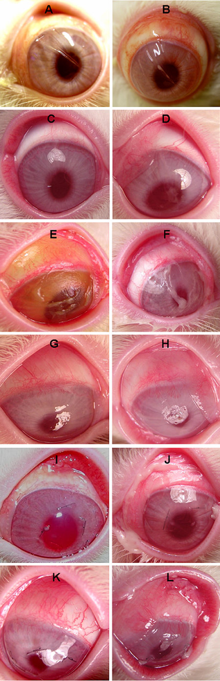
Figure 1. Clinical photos in the saline- or LPS-instilled corneal injury models
Clinical features of the saline-instilled (A,C) or LPS-instilled (B,D) incision model, the saline-instilled (E,G) or LPS-instilled (F,H) scraping model, and the saline-instilled (I,K) or LPS-instilled (J,L) suture model at H4 (A,B,E,F,I,J) and D9 (C,D,G,H,K,L).