![]() Figure 6 of
Kanagavalli, Mol Vis 2007;
13:1161-1168.
Figure 6 of
Kanagavalli, Mol Vis 2007;
13:1161-1168.
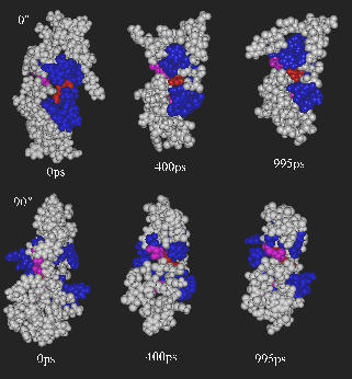
Figure 6. Instability of the arginine mutant structure
Two views of the space filling model at selected time points in the simulation are shown. The position of 367Arg is shown in red. The flanking hydrophobic regions are in blue. The 1ns simulation of the Gly367Arg mutant shows that the burial of the charged residue leads to large fluctuations throughout the model in order to try and accommodate the change. However, large conformation changes are required to prevent the burial of the charged residue.