![]() Figure 4 of
Kanagavalli, Mol Vis 2007;
13:1161-1168.
Figure 4 of
Kanagavalli, Mol Vis 2007;
13:1161-1168.
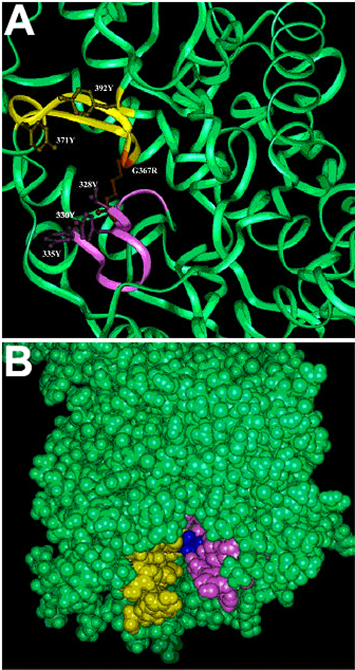
Figure 4. G367R mutation incorporated in the predicted myocilin model showing burial of the mutation in the hydrophobic region and steric hindrance which could lead to the instability and conformational change
A: Steric hindrance and burial of charged residue in Gly367Arg mutant. The ribbon diagram of the mutant region is shown. The mutated residue (gold color) and the neighboring hydrophobic residues (yellow and magenta colors) are shown as ball and stick model. The mutation occurs in a region where two flaps (shown in yellow and magenta) in a cavity (B). B: The mutated region shown in space filling model. The yellow and magenta regions correspond to the flaps shown in A. The side chain of arginine of Gly367Arg (blue) tends to get buried in this hydrophobic region and would therefore lead to instability and conformational change.